
Subdural vs Epidural Hematoma – Key Differences and How to Remember
📅 November 5, 2026 | ⏱️ 10 min read | 📂 Medical
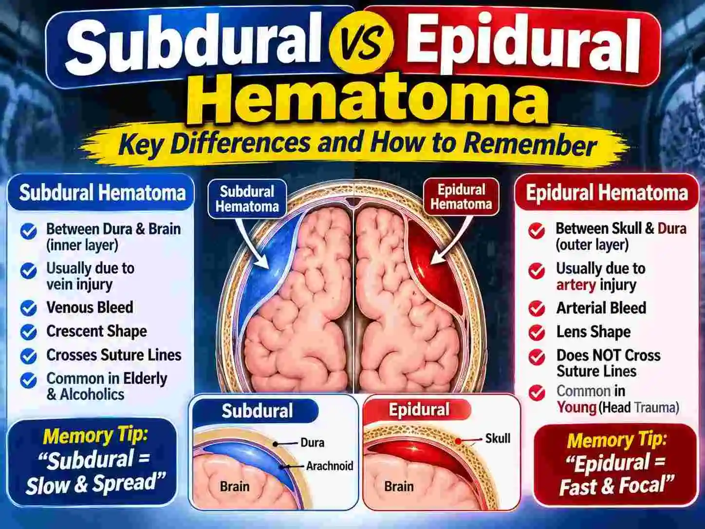
Head trauma is one of the most serious emergencies a medical professional can encounter. Quickly recognising whether a patient has suffered an intracranial haematoma (blood on the brain) is vital. Subdural and Epidural hematomas are two of the most common and dangerous types.
It is essential to know the key differences between an epidural and subdural hematoma. This guide will explain everything including the anatomy, causes, symptoms and treatments of both types. We have also included some useful memory aids to help you.
What is a Hematoma?
Before we compare the two types we need to understand what a hematoma is. A hematoma is blood that has escaped from the blood vessels. If it is located on the brain then blood has escaped and pooled between the brain and the skull. Due to the skull being a rigid, sealed container this puts pressure on the brain which can cause serious damage or death if not dealt with quickly.
Anatomy – Meninges
In order to understand the difference between a subdural and epidural hematoma we need to understand the brain coverings. These are called meninges. Here is the list of layers from outermost to innermost:
- Skull
- Dura Mater (Thick and tough)
- Arachnoid Mater (Middle and web-like)
- Pia Mater (Thin and delicate)
Depending on where the bleeding is occurring will determine the type of hematoma.
Epidural Hematoma (EDH)
An epidural hematoma is when there is bleeding between the skull and the dura mater. The word epidural literally means above the dura.
Causes of Epidural Hematoma
An epidural hematoma is caused by a direct hit to the head causing a fracture to the skull. This usually causes damage to the middle meningeal artery (a large blood vessel) just underneath the skull. As it is an artery the bleeding is fast and at high pressure.
Symptoms of Epidural Hematoma
Common symptoms include:
- Loss of Consciousness
- Lucid Interval (A key sign). Patient will wake up, feel fine but will quickly become worse.
- Headache (Sudden and severe)
- Nausea and Vomiting
- Hemiparesis (One sided weakness) Usually the opposite side to the injury.
- Fixed dilated pupil Usually the same side as the injury.
Subdural Hematoma (SDH)
A subdural hematoma is when there is bleeding between the dura mater and arachnoid mater. This is below the dura.
Causes of Subdural Hematoma
A subdural hematoma is usually caused by a tear in the bridging veins. These are small veins connecting the surface of the brain to the dura mater. Unlike the high pressure arterial bleeding seen with an epidural, subdurals are usually slow and venous.
Often caused by rapid acceleration/deceleration (such as whiplash in a car crash) or minor trauma. Particularly to the elderly or those on blood thinners.
Types of Subdural Hematoma
- Acute SDH – Symptoms occur within 24-48 hours. This is a medical emergency.
- Subacute SDH – Symptoms occur within 2-14 days.
- Chronic SDH – Symptoms occur weeks or even months after the injury. This is very common in the elderly due to the brain shrinking as we age stretching the bridging veins.
Symptoms of Subdural Hematoma
Sometimes the symptoms can be subtle especially if it is chronic.
- Headache (Constant and getting worse)
- Confusion/Memory loss (Common with chronic SDH)
- Personality change (Lethargic/Irritable)
- Weakness/numbness (One sided)
- Seizures
- Loss of consciousness (Usually acute)
Key Differences Between Subdural and Epidural Hematomas
Here is a comparison to fully understand the debate.
| Feature | Epidural Hematoma (EDH) | Subdural Hematoma (SDH) |
|---|---|---|
| Location | Between skull and dura mater. | Between dura mater and arachnoid mater. |
| Cause of Bleeding | Middle meningeal artery (High pressure). | Bridging veins (Low pressure). |
| Typical Cause | Skull fracture. | Acceleration/deceleration (Whiplash), Minor trauma. |
| At Risk Group | Young Adults (Usually trauma). | Elderly, Alcoholics and Blood thinners. |
| Onset | Fast (Minutes to hours). | Slow to Fast (Hours to weeks). |
| Lucid Interval | Very common (Approx 50% of cases). | Uncommon. |
| CT Appearance | Bi-Convex (Lens shape). | Crescent. |
How to Remember Epidural / Subdural Hematomas
Medical students often ask how to remember epidural vs subdural hematoma. The easiest way to remember is using the E and S memory aid.
Epidural (Remember the E):
- Emergency (Fast)
- Early (Onset is early)
- Elliptical (Looks like a lemon on CT scan)
- Extradural (Another name)
- Extremely arterial (Middle Meningeal Artery)
Subdural (Remember the S):
- Slow (Venous so slower)
- Seniors (Most common in the elderly)
- Sickle (Looks like a banana on CT scan)
- Sub-Arachnoid (Located just under the dura).
Diagnosis of Epidural / Subdural Hematomas
If a patient comes into hospital with a head injury this is how doctors will go about diagnosing whether it is an epidural or subdural.
1. Pre-Hospital
First responders will look for signs of trauma and will assess patients consciousness using the Glasgow Coma Scale (GCS). If the GCS has dropped, pupils are unequal or there is a lucid interval then they will be very suspicious of an epidural.
2. CT Scan
A CT scan without contrast is the gold standard for diagnosing intracranial hematomas. It is very quick and accurate.
- Epidural – Looks like a bright white lens shape (Biconvex). Does not cross sutures (Joints between the bones).
- Subdural – Looks like a bright white crescent shape. Can cross sutures but not the midline.
3. MRI
Although a CT scan would be first choice in an emergency sometimes an MRI is used for subacute/chronic subdurals. This gives a clearer picture of the brain tissue and age of the blood.
Treatment
Both require urgent treatment.
Treatments for Epidural Hematoma
As it is usually a fast arterial bleed there is a race against time.
- Surgery – Usually an emergency craniotomy will be required. A Neurosurgeon will remove a section of the skull to relive pressure and stop the bleeding.
- Drugs – Drugs such as Mannitol may be used to reduce swelling of the brain.
Treatments for Subdural Hematoma
Will depend on the size.
- Small Acute/Subacute SDH – Monitored closely in ICU with repeated CT scans.
- Large/Symptomatic SDH – Surgery will be required. Common treatment is a burr hole evacuation. A small hole is drilled into the skull to drain the blood.
- Chronic SDH – Often a simple twist drill procedure can be performed at the bedside to drain the old blood.
Conclusion
Knowing the key differences between a subdural and epidural hematoma is an essential skill to have. An epidural is fast arterial bleed usually caused by a fracture to the skull, whereas a subdural is usually a slower venous bleed commonly seen in the elderly. Using the E/S memory aid and knowing what to look for on a CT scan will ensure you act quickly to save lives.
FAQ
Related Articles
- Glaucoma Symptoms: Early Signs
- Mucocele: Causes, Symptoms and Treatment
- Rule of Nines for Burns
- Winter's Formula Explained